Hemodynamic disturbances of BMD degree ia. What to do if blood circulation between the expectant mother and child is impaired - treatment and preventive measures
Most women do not know about such a test as Doppler until the third trimester, and from that moment Doppler testing becomes a completely common procedure for pregnant women.
Doppler- this is one of the methods ultrasound diagnostics, allowing you to evaluate the intensity of blood flow in various vessels, for example, in the vessels of the uterus and umbilical cord. It is most informative after the 30th week, but if there are deviations during pregnancy (for example, if the fetus is delayed in development), Doppler ultrasound can be prescribed earlier - starting from the 20th week.
Indications for Doppler
Adequate placental blood flow ensures normal pregnancy. Impaired blood flow can lead to delay intrauterine development fetus (IGR), therefore the main reason for prescribing Doppler measurements during pregnancy is precisely the discrepancy between the size of the baby’s body and/or organs and the norms.
It is not necessary that if blood flow is impaired, the child will lag behind in development, but the risk of an unfavorable course of pregnancy increases significantly. Well, and vice versa, if there is a suspicion of a developmental delay in the fetus, but the blood flow is not impaired, then in most cases this indicates that the woman is carrying a low-weight but healthy child.
Doppler ultrasound is also prescribed for:
- premature maturation of the placenta;
- pronounced oligohydramnios or polyhydramnios;
- umbilical cord abnormalities;
- Rhesus conflict;
- gestosis (late toxicosis, complicated by vomiting, severe swelling and increased blood pressure in a pregnant woman);
- the expectant mother has kidney disease, hypertension, diabetes;
- suspected chromosomal pathology;
- non-immune hydrops fetalis;
- uneven development of children with multiple pregnancy(when there is a difference in their body weights of more than 10%).
If the fetus has heart problems, Doppler is performed together with CTG, the so-called Doppler echocardiography.
With feto placental insufficiency Doppler measurements are carried out systematically every 2-3 weeks.
Also, if complications develop during a previous pregnancy, Doppler ultrasound may be prescribed during a subsequent pregnancy.
Preparing for the study and how it is carried out
Doppler testing in pregnant women is carried out according to indications, and mandatory examination at normal course pregnancy is not. But more and more often, in antenatal clinics, all women, without exception, undergo Doppler ultrasound at 30-34 weeks to assess the condition of the fetus.
This procedure is painless and harmless to both mother and fetus. The principle of Doppler testing is the same as a regular ultrasound during pregnancy: a special Doppler sensor is moved across the abdomen, which is equipped with every modern ultrasound diagnostic device. Therefore, this type of research does not require special preparation.
Doppler– this is a visual assessment of blood flow (when an ophthalmologist observes a color and graphic image of blood flow velocity curves from the monitor screen).
Dopplerography- this is the same Doppler, only the readings are additionally recorded on a tape in order to monitor changes (improvement/deterioration) in blood flow after treatment.
Interpretation of Doppler measurements
Uterine arteries (a. uterina dextra - right and a. uterina sinistra - left uterine artery, respectively). The uzist must determine the nature of blood flow in both the left and right uterine arteries, because with gestosis it can be disrupted in only one artery. Thus, by assessing the blood flow in only one artery, one can give a false conclusion, which will negatively affect the health of the baby and the expectant mother.
There is a scientific theory that if blood flow is disrupted in only one (mainly the right) uterine artery, a woman has high risk the appearance of late toxicosis (preeclampsia) with all the negative consequences.
With gestosis, the blood flow in the uterine artery is first disrupted, and as the situation worsens, the blood flow in the umbilical cord arteries deteriorates. Therefore, if blood flow in the uterine arteries is disrupted, it is necessary to periodically repeat Doppler to monitor the situation.
To assess blood flow in the uterine arteries, the resistance index (IR or RI) is calculated.

Often, pregnancy-induced hypertension develops due to impaired uterine blood flow. The expectant mother's body independently increases blood pressure to increase blood flow into the intervillous space. This is how the mother, without realizing it, helps the baby. Thus, it is necessary to improve blood flow and hypertension will disappear on its own.
Impaired blood flow in the uterine arteries is when the value of IR, PI or SDO is greater than normal.
The pulsation index (PI) of the uterine arteries should be within the following limits.

Indicators in the right and left uterine artery may differ slightly from each other. If both indicators are within normal limits, then this picture is not considered a negative phenomenon.
Deviation of blood flow indicators from the norm in two uterine arteries at once indicates a violation of the uteroplacental circulation. This situation requires specific treatment - move more (regularly go swimming or do gymnastics for pregnant women).
Violation of blood flow in only one uterine artery indicates asymmetry of the uterine placental blood flow. If the pregnancy proceeds normally and the baby develops in accordance with the term, then the placenta is fulfilling its functions.
You should be aware that at 18-21 weeks there may be a temporary disruption of blood flow in the uterine arteries. This phenomenon is explained by the fact that the adaptive physiological process Cytotrophoblast invasion has not yet been completely completed. Therefore, if abnormalities are detected in the uterine arteries, a repeat Doppler ultrasound should be performed after 2-3 weeks, i.e. observe the blood flow over time.
The systole-diastolic ratio (SDR) in the uterine arteries should be:

Umbilical cord arteries (a. umbilicalis). To obtain true results, the study should be carried out only while the baby is at rest, and only when his heart rate is between 120-160 beats per minute. After all, physiologically it is so laid down that when the heart rate increases, the IR in the umbilical cord artery decreases, and vice versa, when the heart rate decreases, the IR increases.
Measuring blood flow in the umbilical cord arteries should be done while the pregnant woman is lying on her back! Assessment of the severity of umbilical cord blood flow disturbance cannot be objective based on the location expectant mother"on the left side."
The umbilical cord should have two arteries and one vein. If there is an anomaly (a single umbilical cord artery), then the fetus may suffer from a lack of oxygen and nutrients, which is why it lags behind in weight and growth. But it happens that the fetus adapts to such an existence and does not experience a deficiency necessary substances. Such babies are born with low weight, but absolutely viable. Therefore, if there is one umbilical cord artery and the blood flow in it is not impaired, then there is no cause for concern. But if the blood flow in a single artery is disrupted, a hospital treatment to improve blood flow and, if necessary, early delivery (if the fetus is severely delayed in development).
The most widely used method for assessing the nature of blood flow in the umbilical cord arteries is the resistance index. The readings in both umbilical cord arteries should be almost the same.
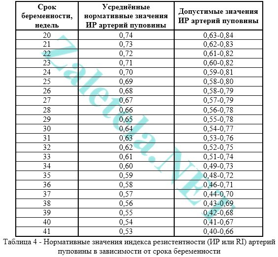
Impaired blood flow in the umbilical cord is when the value of IR, PI or SDO in the umbilical cord arteries is higher than normal.
The pulsatility index (PI or PI) of the umbilical cord arteries must meet the following standards:

Registration of zero and reverse values of diastolic blood flow is pathological. This means that the fetus is in critical condition.
There are only 2-3 days left from the moment constant reverse values appear until the death of the fetus, so it is necessary to carry out C-section to save the baby's life. This is only possible starting from week 28, when the baby is viable.
Systole-diastolic ratio (SDR) in the umbilical cord arteries:

If the blood flow in the umbilical cord is impaired, then, as a rule, fetal development is delayed. If there is no developmental delay now, but the blood flow in the umbilical cord is impaired, then without treatment, the fetus may experience developmental delay.
Average cerebral artery fetus (a. cerebri media). When the fetus suffers, it is observed increase in the values of PI, SDO and speed in the SMA.

Maximum velocity (aka V max) in the fetal middle cerebral artery:

Systole-diastolic ratio (SDR) for the middle cerebral artery:

Fetal aorta. It leaves the left ventricle of the heart, runs along the spine and ends at lower area abdomen, where the aorta divides into two iliac arteries, which provide blood supply to the human legs.
Abnormalities in the blood flow of the aorta can only be detected after 22-24 weeks of pregnancy.
Impaired blood flow is increasing the values of IR, PI and SDO. Critical (indicating fetal death) is considered registration of extremely low values until their complete disappearance.

Changes in the aorta characterize the severity intrauterine hypoxia fetus


Systole-diastolic ratio (SDR) for the fetal aorta:

Ductus venosus (DV). It is studied with enhanced Doppler assessment of blood flow.
During the study, it is necessary not to take into account episodes of hiccup-like respiratory movements of the child and active movement.
Indices are not used to assess the ductus venosus.
Diagnostic criterion pathological condition fetus is considered to be present negative or zero blood flow values during the phase of atrial contraction. Zero or reverse values are recorded for fetal malnutrition, congenital defects of the right heart, and non-immune fetal hydrops.
Even with critical blood flow in the umbilical cord arteries, but with preserved blood flow in the ductus venosus during the phase of atrial contraction, it is possible to extend gestation to optimal timing for childbirth.
Description of blood flow disorders and their treatment
1st degree
1 A degree– disturbance of blood flow in the uterine arteries, while in the umbilical cord blood flow remains normal.
This degree of blood flow disturbance is not dangerous for the fetus.
Drug treatment for this condition is ineffective. Doctors still prescribe therapy with Actovegin and Curantil. Don't see each other on occasion!
In fact, if blood flow in the uterine arteries is impaired, it is more advisable to simply walk more often. fresh air(breathing full breasts) + eat right + move more (walking, special exercises for pregnant women, morning exercises, yoga, swimming). And don't sit at the computer for hours! That's all the treatment is.
1 B degree– disturbance of blood flow in the umbilical cord arteries, but hemodynamics in the uterine arteries are normal.
This degree of blood flow impairment requires the use of blood thinning drugs to avoid developmental delay and fetal hypoxia.
In this case, treatment is prescribed aimed at improving blood circulation (the drug Placenta compositum, Curantil or Trental). Actovegin is prescribed as an antihypoxant that improves oxygen supply to the fetus.
A blood test for clotting ability (coagulogram) is also prescribed. In case of increased blood clotting, it is necessary to take stronger blood-thinning drugs than Curantil (for example, heparin or a product containing acetylsalicylic acid).
Degree I of the disorder does not lead to fetal death. Systematic monitoring of the nature of blood flow is carried out (every 2 weeks) “plus” monitoring of fetal CTG (after 28 weeks of pregnancy). In addition, be sure to monitor the blood pressure of a pregnant woman.
2nd degree– simultaneous disturbance of blood flow in the uterine arteries and in the umbilical cord, which does not reach critical values (when blood flow is preserved in the venous duct).
In this condition in mandatory drug treatment is prescribed in a hospital, where round-the-clock monitoring of the fetal condition is provided. It is also necessary to monitor the state of blood flow by performing Doppler + CTG every 2 days.
In grade II, hemodynamic disturbances are rare, but cases of intrauterine mortality may occur.
3rd degree– critical disturbances of blood flow in the umbilical cord with intact or impaired blood flow in the uterine arteries. A critical violation is understood as registration of reverse diastolic blood flow or its absence at all.
The third degree of violation poses a danger to the child’s health, because in half of the cases the baby’s intrauterine death occurs. Therefore, if a 3rd degree of blood flow disorder is detected, it is necessary to urgently perform a cesarean section in order to save the baby’s life, because at this stage of the disorder, treatment is not effective.
Conservative (natural) childbirth in the 3rd degree can lead to perinatal death of the child.
Cost of Doppler ultrasound in private clinic– about 1,200 rub.
There are many reasons that contribute to impaired blood flow during pregnancy. Let's consider the most common factors that provoke blood flow disorders.
- Diseases of the uterus: bicornuate uterus, endometriosis, uterine hypoplasia, presence of fibroids, etc.
- Maternal health problems: renal failure, diabetes mellitus, hypotension, pyelonephritis, endocrine system diseases, bronchial asthma and etc.
- Unfavorable gestation conditions: Rh-conflict, multiple births, gestosis, malpresentation of the fetus, etc.
- External factors: drinking alcohol during pregnancy, smoking, constantly being in a nervous environment, first birth (and the woman is over 35), poor (limited) maternal nutrition.
Classification of blood flow disorders in the placenta
Placental insufficiency negatively affects the functioning of the placenta. It can be acute or chronic.
Clinical symptoms of PN depend on the nature of the pathology. Acute form deficiency can develop in any trimester of pregnancy.
There is a disruption in the gas exchange function of the placenta, which can lead to fetal hypoxia. The acute form develops due to premature placental abruption or vascular thrombosis.
Causes of uterine blood flow disorders
The causes of impaired blood flow during pregnancy can be found in the following pathological conditions:

- Anemia (anemia) of the expectant mother. Low hemoglobin levels cause high blood flow rates. This occurs by compensating for the lack of oxygen and cannot but affect the processes in the development of the fetus and the quality of blood exchange;
- The nature of the position of the placenta. If presentation is diagnosed, which can be justified by a previous caesarean section, then the blood supply will definitely be reduced due to thinning of the uterus at the site of the scar;
- Late toxicosis causing pathological changes in the work of small vessels. This is one of the most common signs of blood flow disorders during pregnancy;
- Viruses and infections present in the mother’s body during gestation. Some of them can cause damage to placental tissue and contribute to the development of placental insufficiency;
- Rhesus conflict - may be complicated by the anemic condition of the fetus;
- Fluctuating blood pressure levels that do not allow maintaining uniform blood flow rates;
- Defects of the uterine organ. The most significant of them, which can change the course of fetal development for the worse, is the two-cavity structure of the uterine sac. The uterine space, divided into two parts, is not in itself an obstacle to normal height and formation of the child. However, the blood supply system does not adequately supply such a two-chamber cavity;
- Serious quantitative or configurational changes in the vessels of the umbilical cord;
- Damage to the inner wall of the uterus resulting from surgical interventions, or as a consequence of bad habits;
- Tumors, such as fibroids, are especially dangerous in nulliparous women after thirty-five years of age. This also applies to uterine fibroids, which are abundantly saturated with blood during perinatation. Against the background of an increase in size and formation of the myomatous node, a persistent lack of blood flow to the placenta is formed;
- Pregnancy complicated by multiple pregnancy. Since the placenta is forced to adapt to maintaining several feeding organisms in proper conditions at once, errors in the blood supply such as the donor position of one of the fetuses cannot be ruled out. Often, there is underdevelopment of the feeding fetus, a significant lack of weight and physiological normal signs. The child, involuntarily acting as a recipient, also seriously suffers from, on the contrary, an overly abundant blood supply;
- The mother's diabetes, sometimes developing precisely during pregnancy, loosens the walls of blood vessels, which does not have the best effect on blood circulation.
Poor blood circulation in the uterus can be caused by increased pressure, pneumonia, intrauterine infection and insufficient oxygen supply to the fetus (hypoxia).
To diagnose the blood flow system in obstetric practice, three-dimensional ultrasonography(Doppler), with the help of which the vessels are visible in the so-called 3D (three-dimensional) image.
With the help of this modern diagnostic method, there is a prospect of diagnosing retroplacental bleeding and assessing cardiac malformations by monitoring blood flow.
This method is indispensable, since with its help you can see defects even in the smallest vessels that form the microvasculature, monitor the development and formation of intraplacental hemodynamics, and also control the amount of oxygen and nutrients that should enter the fetal body.
New opportunities have opened up for early detection obstetric complications, and if correction or treatment is started without wasting time, then circulatory disorders and further associated pathologies can be practically avoided.
Symptoms of pathology
Like any other pathology, BMD disorder has a number of features in its manifestation. If you know exactly the signs of this deviation, a woman will be able to identify the disease in the early stages, which will allow her to consult a doctor in time. The main danger of stage 1a uteroplacental blood flow disturbance is that the fetus experiences oxygen starvation. Such hypoxia prevents the normal development of its internal organs, can cause miscarriage or fading of pregnancy. Pathology can be recognized by the following changes:

- the child's heart rate increases significantly;
- the fetus periodically becomes either active or lethargic;
- the volume of the abdomen does not correspond to normal readings - it is ahead of them.
Signs of degree 1a BMD impairment usually appear in the decompensated form. However, in some cases there are no manifestations of this pathology at all. It is possible to find out about its presence only after the next examination.
Manifestations of FPN depend on their type. With compensated chronic fetoplacental insufficiency, there are no symptoms. A woman learns about abnormalities during an ultrasound examination.
Acute and chronic decompensated forms of pathology are characterized by severe symptoms. A woman may notice periods of vigorous motor activity of the unborn child, which are followed by periods of complete rest.
There are certain standards, according to which a pregnant woman over 28 weeks should feel at least 10 fetal movements per day. With more low rates a woman should consult a gynecologist.
Additional signs of impaired blood flow may be a slowdown in the increase in abdominal circumference. It is difficult to identify this on your own, so you need to visit regularly antenatal clinic, where such measurements are regularly carried out.
The most dangerous symptom of FPN is bloody issues from the vagina. This may be a sign of placental abruption. This condition requires urgent medical attention.
Diagnosis of pathology
It was previously said that during pregnancy, blood flow disorders can be diagnosed using Doppler ultrasound. It is an ultrasound examination that can detect any pathological abnormalities in blood flow. When diagnosed, a pregnant woman takes a horizontal position on her back or side. The specialist conducts the examination using the transabdominal method. Usually Doppler testing is prescribed twice:
- at 20–22 weeks, to ensure that there are no abnormalities in the development of the fetus;
- at 32 weeks.
Identification of pathological disorders of placental blood flow is carried out through a comprehensive examination, but ultrasound, which is combined with Doppler measurements, plays a huge role in diagnosis. This method allows us to identify not only blood flow disorders, but also complications caused by them.

Doppler measurements are prescribed in the following cases:
- diseases of the mother that can provoke disturbances in blood flow in the placenta;
- premature aging placenta;
- intrauterine growth retardation syndrome;
- polyhydramnios or oligohydramnios;
- signs of fetal hypoxia;
- birth defects And genetic diseases in the fetus.
Depending on complexity pathological process, disturbances can be observed in the umbilical, uterine or fetal vessels. Based on the results of the examination, a diagnosis of uteroplacental, placental or fetoplacental form of blood flow disturbance is made.
Atypical blood circulation in the placenta may be indicated by: indirect sign, such as its thinning or increase in area, symptoms of intrauterine infection and changes amniotic fluid.
The main diagnosis of blood flow disorders during pregnancy is this moment, is a Doppler examination. The most insignificant, at first glance, changes in one of the early stages of disorders are clearly visible on the screen, since Doppler imaging provides a picture in color and has a high degree of resolution in terms of picture clarity.

The very analysis of the frequency of ultrasonic waves reflected from objects in motion makes it possible to determine the speed of blood flow through the arteries, in the umbilical vessels, as well as blood circulation inside the placenta.
Then the indicators taken during the examination are checked against the table of normative data and the result is recorded.
The biggest advantage of Doppler ultrasound, in addition to the safety of the study, is the accurate prediction of any form of abnormality, detecting them at a stage when it is still possible to save the fetus and not cause harm to the mother’s body.
In addition to ultrasound examination, classical measures to confirm or refute pathology are mandatory:
- Analysis of fetal activity based on the mother’s words;
- Stethoscopic listening to the baby's heartbeat in the womb;
- Cardiac examination.
Treatment methods
If the disease occurs in mild form(first degree), then the doctor may prescribe medications that improve blood circulation.
The dynamics of the fetal condition are carried out; weekly, until the indicators normalize, the pregnant woman undergoes Doppler measurements and checks the fetal heartbeat. If the indicators stabilize, the woman will continue to bear the child.
In the second degree, the pregnant woman is hospitalized and treated under the strict supervision of medical staff. If the condition worsens, an unscheduled operation is performed.
As for the third degree, it cannot be treated, since irreversible changes begin in the development of the fetus. Therefore, in order not to risk the child’s life, doctors insist on an immediate cesarean section.
It is impossible to treat disorders of the uteroplacental blood flow, acting only in one direction, or eliminating problems as they arise.

A complete cure necessarily includes a set of measures aimed at:
- Increased blood microcirculation;
- Achieving optimal blood pressure;
- Vasodilation with spasmodic manifestations in the arteries;
- Reducing uterine tone due to relaxation of blood vessels;
- Preventing the consequences of oxygen starvation (hypoxia);
- Saturation of placental tissue with the phospholipids it needs.
In conclusion about prevention
Prevention actions should be aimed at organizing conditions for the healthy growth and intrauterine development of the child. To do this, a woman must:
- watch your diet;
- rest more often;
- regularly spend time in the fresh air;
- to refuse from bad habits;
- minimize emotional stress.
The main thing is to regularly visit your doctor and follow his recommendations.
To give birth healthy baby, a pregnant woman needs to be more careful and listen to her body. Try to watch your diet: it should be nutritious, rich and healthy.
You can also take vitamin complexes or Ginipral, which will eliminate the deficiency of any element. Also try to drink as much clean water as possible - at least 2 liters per day.
Do not forget to control your body weight - during pregnancy it should not increase by more than 10 kg.

If normal blood flow is disrupted, the woman may be placed on preservation. Remember that you should not prescribe treatment for yourself based on the advice of friends or information from the Internet.
This will lead to the development of serious complications.
Preventive actions aimed at timely identification of risk groups among pregnant women. Currently, there is no uniform treatment method for this condition. As a rule, therapy is complex and aimed at stabilizing the condition in order to avoid premature birth.
Doctors recommend sleeping on the left side; medications containing amino acids, ATP, and glucose are often prescribed to stabilize metabolic processes. May also be recommended medicines, reducing the tone of the uterus, normalizing blood circulation, vasodilators and drugs that reduce blood clotting.
Only a doctor can prescribe treatment; sometimes hospitalization in a hospital is required for a full examination, monitoring and therapy. If there is a significant deterioration in placental blood flow, an emergency caesarean section is prescribed.
In order not to encounter placental insufficiency during the period of bearing a child, it is necessary to reconsider your lifestyle even during pregnancy planning and eliminate all possible risks.
Every woman who wants to give birth to a child must remember that the mother’s condition is completely transmitted to the unborn baby. Therefore, in order for the fetus to develop without complications, she needs to make up her diet from food containing a maximum of vitamins, micro- and macroelements, as well as rich in the required amount of carbohydrates, proteins and fats.
If a pregnant woman is not bothered by swelling, then fluid intake should be at least 1-1.5 liters.
It is important to monitor changes in body weight, since by the end of pregnancy the weight gain should not exceed 10 kg.
There are risk groups that require the use of drug prophylaxis, which promotes the interaction of the body systems of the fetus and mother and prevents dysfunction of the uteroplacental circulation.
Timely corrected methods of labor management will help to significantly reduce perinatal morbidity and mortality. drug therapy. But a high risk of severe neurological complications cannot be ruled out.
Especially for beremennost.net Elena Zhirko
In the process of bearing a child, a woman's body inevitably changes. Since impaired blood flow during pregnancy occupies one of the leading positions among all pathologies of the gestational period, assessment of the state of blood circulation between the expectant mother and baby is included in the mandatory examination program for pregnant patients.
Why does disruption of the uteroplacental blood flow (UPB) occur? What types of this pathological process exist? What is the difference between grades 1a and 1b? How dangerous is this phenomenon for a child? What to do if blood flow is impaired? In what ways is its condition checked?
Degrees of disturbance of uteroplacental blood flow
The uteroplacental blood flow is an anatomically complex system consisting of the placenta, blood vessels of the expectant mother and the child. Disorders of uteroplacental blood flow (UPF) are common pathologies caused by dysfunction of the placenta and umbilical cord.
When diagnosing this pathological phenomenon, grades 1, 2 and 3 are distinguished. In this case, the first degree is divided into 2 types. Information about each of them is presented in the table.
| NPMK degrees | Characteristic | Possible consequences | |
| 1 | 1a | Poor communication between the uterus and the placenta when the latter is fully connected to the embryo. | Deviations in the development of a child in mild forms, manifested in the form of underweight and disturbances in general physical characteristics. |
| 1b | The state of the uteroplacental blood flow is normal, but the embryo-placenta blood circulation pattern has deviations. | Developmental delay. | |
| 2 | Placental insufficiency is present at each level. It is almost impossible to compensate for the lack of oxygen, because The embryonic aorta, uterine artery and umbilical cord vessel are not able to fully pass blood. | In 85% of cases the child dies. | |
| 3 | Characterized by centralization of blood flow. | The condition of the fetus becomes critical due to dysfunction of intracardiac hemodynamics. During Doppler measurements, reverse diastolic blood flow is often recorded. The fetus has developmental abnormalities. This degree can rarely be cured. | |
Pathologies are also classified according to other criteria. The table shows the types of disease.
| Sign | View | Description |
| By time of occurrence | Primary placental insufficiency | Develops before 16 weeks of pregnancy. It manifests itself in the form of disruption of the process of embryo attachment and further abnormal formation of the placenta. |
| Secondary placental insufficiency | It is detected by the time the placenta is already fully formed - after 16 weeks of gestation. Pathology occurs under negative impact external factors. | |
| According to symptoms | Compensation | Metabolic disturbances occur in the functioning of the placenta, but the blood flow between this organ and the uterus or fetus functions without interruption. |
| Subcompensation | The female body is not able to restore the blood supply to the embryo necessary for its full growth, because all elements of the blood flow system are not working properly. | |
| Decompensation | There is a disruption of blood flow at all levels, which is difficult to treat. |

Causes that can lead to pathology
Many factors contribute to the appearance of disturbances in uterine blood flow. Many of them are capable of influencing the placenta not only at the stage of its formation, but also at later stages. Possible causes of deterioration of uteroplacental circulation:
- Anemia. Due to a decrease in the concentration of hemoglobin in the blood, hemodynamic parameters increase in all blood vessels, including the uterine ones. This is due to the fact that the body seeks to restore the supply of oxygen to tissues by increasing the speed of blood flow, including uterine.
- Incorrect attachment of the placenta. Accompanied by a decrease in blood flow due to thin muscles in the lower segment of the uterus. This problem occurs when the placenta is attached to its scarred area. This zone cannot provide uteroplacental blood circulation, as a result of which the blood supplied to the embryo may not be enough for full intrauterine development.
- Late toxicosis. This condition, during which small blood vessels are affected, often provokes a violation of the uteroplacental-fetal blood flow (UPFB).
- Infectious diseases suffered by a woman during gestation. A number of pathogenic agents negatively affect the condition of the placenta, causing pathological changes in its tissue. Consequence - the IPC was violated.
- Conflict between the Rh factors of the woman and the fetus. This leads to the development of anemia in the baby, which is fraught with a deterioration in the blood supply to his body.

- Pressure surges. They negatively affect blood circulation in the vessels, provoking the development of NMPK.
- Abnormal structure of the uterus. The bicornuate organ has a septum. Pregnancy develops in one of the two cavities formed. The danger in this case lies in the disruption of the child’s full blood supply. Normally, this is provided by two uterine arteries. During gestation, their diameter increases, which leads to the formation large number the vessels connecting them, which help normalize blood flow. In a uterus with such an abnormal structure, these processes are absent, so the required volume of blood does not reach the placenta.
- Defects of the umbilical cord vessels. When their number changes, NMPC develops.
- Endometrial pathologies. Their development is caused by inflammation, surgical interventions, and bad habits of the expectant mother.
- Myoma. With the development of neoplasms, their blood supply increases, and the flow of blood to the fetus, on the contrary, decreases.
- Multiple pregnancy. When implanting two or more fertilized eggs the placental area increases significantly. In addition, it is possible for a larger volume of blood flow to transfer to one of the embryos. Not only the donor child suffers, but also the recipient fetus, because his heart muscle is not ready for such an amount of incoming blood.
- Diabetes. By affecting the internal walls of the arteries, this pathology triggers the development of placental insufficiency.

How dangerous is a 1st degree violation for a child?
The most common and dangerous consequence These hemodynamic disorders (HDN) are oxygen starvation. Other complications of poor blood supply to the fetus include:
- decrease in body weight and physical parameters (intrauterine growth retardation);
- acid-base imbalance;
- disorder of the heart in the form of acceleration or deceleration of the pulse, arrhythmia;
- reduction of adipose tissue in the body;
- threat of pathological abortion;
- hormone imbalance;
- antenatal fetal death.

Symptoms of uteroplacental blood flow disorder 1a degree
If this pathology is in the stage of compensation, the expectant mother will not feel any pronounced deviations. About the disease in in this case can be found out only after the examination. Clear signs diseases accompany the acute form and chronic decompensation. This pathology is accompanied by the following symptoms:
- a sharp increase or cessation of motor activity of the embryo;
- too much slow growth abdomen (the diameter of its circumference does not correspond to the standard indicators corresponding to specific date gestation);
- gestosis;
- arterial hypertension;
- strong weight gain of the expectant mother;
- swelling of the legs below the knees;
- proteinuria.

In some cases there may be bleeding. This symptom most likely indicates placental abruption. If bleeding occurs, you should immediately see a gynecologist.
Diagnostic methods
Dopplerography can provide the most reliable and complete information about this pathology. This diagnostic procedure is based on the use of ultrasonic waves and is completely safe for the expectant mother and baby. Using the procedure, signs of circulatory disorders are diagnosed, such as a decrease in diastolic velocity, an increase in the resistance index, and a dicrotic notch in the blood flow curve. The table provides information on how this pathology is diagnosed.
| Diagnostic method | Type of study | Purpose of the event |
| History taking | Analysis of the patient’s complaints, correlation of abdominal circumference with standard indicators corresponding to the gestational age | Making a preliminary diagnosis, developing a plan for further action |
| Physical examination | Auscultation | Definition heart rate fetus |
| Laboratory research | Blood analysis | Determination of the amount of estrogens, progesterone, human chorionic gonadotropin |
| Instrumental studies | Ultrasound of the pelvic and abdominal organs | Determining the size of the fetus and the condition of the placenta |
| Cardiotocography | Study of the child’s heart function | |
| Dopplerography | Assessment of the intensity of blood flow, determination of the state of intraplacental circulation, flow speed and direction of blood in the vessels of the uterus and umbilical cord |

Features of treatment during pregnancy
Therapeutic tactics depend on the degree of the pathological process and the pathogenesis of the disorders. Treat medications This disease is possible only with the first degree of circulatory impairment. The second degree is considered borderline. If the pathology has reached the third degree, it is indicated surgical intervention. The doctor decides which treatment method to choose on an individual basis.
Conservative methods of therapy
Therapeutic tactics are based on a complex effect on all elements of the hemodynamic process:
- For minor deviations from the norm, Hofitol is used. If symptoms are severe, the patient is prescribed drugs with more active ingredients (Pentoxipharm, Actovegin) (see also:).
- When a pregnant woman is diagnosed with a tendency to form blood clots, medications are used that can improve the flow of blood through the blood vessels (Curantil).
- To dilate blood vessels, Drotaverine or No-Shpa is used orally, Eufillin is used as injections.
- For uterine hypertonicity, drip administration of magnesia and enteral use of Magne B6 are indicated.
- The negative consequences of circulatory disorders must be eliminated with the help of ascorbic acid and tocopherol, which have an antioxidant effect.

Medicines are prescribed by the attending physician. Self-medication is strictly prohibited. If the chosen treatment tactics do not improve well-being, the patient is indicated for inpatient treatment. This measure will allow for constant medical monitoring of the condition of the expectant mother and fetus.
Surgical intervention
If signs of pathology are pronounced (grades 2 and 3 MPC), emergency delivery is resorted to. In situations where conservative therapy did not give the expected result, including that which was carried out with diagnosed 1st degree of blood flow impairment, a decision on further actions is made in the next 48 hours. In this case, as a rule, doctors perform a caesarean section. If childbirth in this way is planned to take place before 32 weeks of gestation, the baby’s condition and vital signs must be assessed.
In the opposite direction, unnecessary substances formed as a result of biochemical processes are removed.
Impaired uteroplacental blood flow causes a condition called placental insufficiency. This leads to fetal death and miscarriage.
For 36 weeks, three mandatory ultrasound examinations are performed. It allows you to timely identify the disorder, develop a plan for managing pregnancy and childbirth, prescribe treatment, prevent death and abnormal development child.
Modern requirements of obstetricians and gynecologists are aimed at examining pregnant women using safe methods to assess uteroplacental blood flow by volume.
How does blood circulation function between mother and fetus?
The mother-fetus circulatory system is based on such anatomical structures as the placenta, umbilical arteries, and veins.
Blood enters the placenta through the uterine arteries. The structure of their walls is distinguished by the presence of a muscle layer that can contract and block the lumen. Before pregnancy occurs, this mechanism helps reduce blood loss during menstruation.
At 4–5 weeks of consolidation of the fertilized egg (gestation process), the muscle layer disappears. Blood flow to the placenta no longer depends on vascular contraction. And by the sixteenth week, the arteries are transformed for constant blood supply. This turns out to be dangerous when bleeding occurs, since it is impossible to stop it by reducing the lumen of the vessels.
What happens here at the cellular level:
- exchange between the maternal body and the fetal bloodstream;
- two differently directed flows meet;
- the transfer of necessary substances takes place (diffusion).
The other part of the general blood circulation is provided by the vessels of the umbilical cord (normally there are 2 arteries and a vein). The main volume of blood flows to the fetus through the arteries, and flows through the veins towards the placenta.
As the uterus grows, the arteries expand and form anastomoses.
Violation of fetal-placental blood flow is most difficult to tolerate developing child. Creates conditions for an unsatisfactory prognosis for the development of internal organs and systems and the birth of a healthy baby.
What reasons can break the blood flow between the mother, placenta and fetus?
The causes of disruption of the circulatory system between the maternal body and the fetus (fetoplacental insufficiency) have been well studied. Some factors are formed only during pregnancy. The other depends on the general health of the woman.
Pregnancy pathologies include:
- Low attachment of the placenta (obstetricians say - presentation, “placentation”) - the lower parts of the uterus are distinguished by a thinner muscle layer. Through it, not enough blood flows to the fetus. A similar situation develops in the case of presentation in the area of a postoperative scar (for example, from a cesarean section).
- Late toxicosis is accompanied by damage to small vessels of the uterus; the complication is the most common blood flow disorder.
- Anemia - low level hemoglobin causes a compensatory acceleration of the heartbeat, and blood flow through the uterine arteries increases to compensate for the lack of oxygen. Circulation also changes in the uteroplacental circle.
- Incompatibility between the blood of mother and fetus according to Rh - an immune conflict arises with the development hemolytic disease child, anemia. The same situation is possible when transfusion of different blood types from a donor.
- The load on the kidneys due to toxicosis can cause an increase in blood pressure. This helps change blood flow.
- Pathology of the umbilical cord arteries is rarely detected. If there is only one umbilical artery, then there is insufficient blood flow to the fetus.
- Multiple pregnancy - the placenta is increased in size and requires increased nutrition. Sometimes blood flow changes from one fetus to another.
It turns out that the first child is a constant donor for the twin, develops worse, because he transfers blood to his brother, and he himself is “malnourished”
Such changes are called fetotransfusion syndrome. The donor has a lower body weight. And the recipient experiences an increased load on the developing heart. Both kids have problems.
The most dangerous diseases for women are:
- Acute infections during pregnancy - pathogens can penetrate the placental barrier and destroy the vascular network.
- Malformations of the uterus - the most significant is the “bicornuate” uterus. Inside the cavity there is a partition dividing it into 2 parts. Pregnancy is possible only in one of them. The main violation is not the compression factor (the cavity has the ability to stretch sufficiently), but the lack of communication between the uterine arteries, insufficient development of the vascular network, and placental hypoxia.
- Endometriosis - changes in the inner lining of the uterus that occur after inflammatory diseases(including sexually transmitted infections), frequent abortions, diagnostic curettages. One of the reasons is smoking and alcohol.
- Tumor of the uterus - if a woman has even a small fibroid ( benign tumor), then pregnancy stimulates the growth of nodes. They take over part of the blood supply, and the fetal blood flow is “robbed.” Failure directly depends on the size of the tumor.
- Diabetes mellitus - affects the walls of blood vessels, often occurs in women with risk factors during pregnancy.
How does insufficient placental blood supply threaten the fetus?
All disorders of both uteroplacental and fetal placental nature lead to oxygen deficiency fetus (hypoxia). Complications are caused precisely by this mechanism:
- the formation of the internal organs of the fetus is disrupted, there is a lack of weight, this is called “intrauterine growth retardation”;
- the heart reacts with rapid contractions (tachycardia) or arrhythmias, bradycardia;
- the composition of electrolytes and acid-base balance are disrupted;
- the functioning of the endocrine system is disrupted, the fetus experiences a hormonal imbalance;
- fat depots are not formed.
The most severe complications are fetal death and threatened miscarriage.
Myomatous nodes take away part of the vascular network from the fetus for its growth
Types of blood flow disorders in the placenta
There are fetoplacental (between the fetus and placenta) insufficiency and uteroplacental insufficiency.
Fetoplacental hypoxia can occur as:
- Acute deficiency - occurs during any period of pregnancy and during labor. Causes premature placental abruption, vascular thrombosis, infarction in the placenta area, and hemorrhages. Capable of causing the death of a child.
- Chronic - occurs more often, develops from the second trimester, but manifests itself only in the third. Changes in the placenta are in the nature of premature aging; fibrin is deposited on the surface of the villi. Permeability is sharply reduced, which provokes fetal hypoxia.
Against the background of the development of chronic placental insufficiency, the following stages can be distinguished:
- compensation - the course is favorable, since the protective mechanisms of the mother’s body are triggered and compensate for the baby’s missing nutrition, the treatment is effective, the child is born on time, healthy;
- subcompensation - the mother’s body is not able to fully compensate for the “unprofitable” blood supply to the fetus, full treatment is necessary, the child may be born with complications and lags behind in development;
- decompensation - pathology develops rapidly, compensatory mechanisms are insufficient, the fetus’s heart activity is disrupted, intrauterine death is possible;
- critical stage - characterized by pronounced structural changes in the placenta, which disrupts its functions, therapy cannot change the condition of the fetus, death is inevitable.
Degrees of impaired blood flow
In the joint violation of fetoplacental and uteroplacental blood flow, 3 degrees are distinguished.
I - changes are compensated, do not threaten the fetus, affect only the uteroplacental blood flow, the child develops normally. Depending on the level of changes, there are:
- degree Ia - disturbance of uteroplacental blood flow is limited to one of the uterine arteries, all hemodynamic parameters are stable, within normal limits;
- degree Ib - blood flow is disrupted at the level of communication between the fetus and the placenta due to the vessels of the umbilical cord; enough blood flows through the uterine arteries.
If minor changes in the first stage were not detected and the woman did not receive treatment, then after 3-4 weeks, second-degree disorders occur.
II - blood flow in the uterine and umbilical arteries changes.
III - indicators are critical, reverse blood flow in the arteries is possible.
How is diagnosis carried out?
Using Dopplerography, it is possible to examine blood flow through arteries and veins, obtain a color graphic image, and measure fetal hemodynamics.
This plays a significant role in predicting the course of pregnancy and creates conditions for making decisions on treatment measures.
Indirect diagnostic methods include:
The methods allow us to identify lack of fetal weight and placental dysfunction. These signs may be evidence of the development of hypoxia.
What does the mother feel and what does the doctor determine during the examination?
Hypoxia stimulates motor activity fetus
At an appointment with an obstetrician-gynecologist, the doctor listens to the fetal heartbeat and pays attention to high frequency, arrhythmia or bradycardia. This necessitates referral for Doppler examination.
A pregnant woman pays attention to increased movements, tremors
Treatment of disorders
Establishing the degree of impaired uteroplacental blood flow is necessary for choosing pregnancy management tactics.
- It is believed that it is possible to maintain a pregnancy in the first degree (a and b); treatment will also help.
- The second degree is considered borderline, requiring constant monitoring; the effectiveness of treatment is unlikely.
- In the third degree, urgent delivery using surgical methods is required.
Treatment options are aimed at all parts of the pathology:
- to improve microcirculation, use Pentoxifylline, Actovegin;
- to support low blood flow speed and pressure in the vessels, Stabizol, Venofundin, Infucol are used (synthesized on the basis of a starch solution, capable of retaining fluid in the vessels);
- vasodilating drugs such as Eufillin, No-shpa eliminate spasm of medium and small arteries;
- by reducing the tone of the uterus, it is possible to influence vascular spasm, reduce the degree of hypoxia, use magnesium sulfate, Magne B6, Ginipral;
- antioxidants eliminate the effects of hypoxia, destroy decay products, prescribe Tocopherol, combinations of vitamin E and ascorbic acid, Chophytol;
- Essentiale has a protective effect by increasing the level of beneficial phospholipids in the blood and improving liver function;
- Curantil is prescribed during pregnancy against the background of uterine fibroids; a positive effect on microcirculation and the prevention of thrombosis has been established.
Obstetricians continue to use Cocarboxylase in practice, which cardiologists have abandoned. But gynecologists consider the drug effective for restoring tissue respiration.
Incubators are used for the treatment and care of newborns as indicated.
Forecast and consequences
For statistical studies, an indicator such as “perinatal mortality” is used. It includes all deaths occurring in a fetus from the 22nd week of pregnancy and among newborns in the first week of life. It is believed that it fully reflects the influence of the factor of pregnancy and childbirth. The calculation is per 1000 children born.
Currently, 13.3% of children die from the second degree of disturbance of the uteroplacental circulation, and up to 47% in the third degree. Timely caesarean section reduces mortality.
Intensive care needs:
- 35.5% of newborns with the first degree;
- 45.5% – from the second;
- 88.2% - from the third.
The consequences of preserving and treating children born in conditions of pathological hypoxia are unclear. Pediatricians and psychiatrists point to its unconditional influence on physical and mental development.
Only experienced specialists can diagnose and treat conditions associated with disruption of the uteroplacental barrier. You should not take medications on your own or take advice from uneducated people. The situation can become critical not only for the fetus, but also for the woman.
I was told the uteroplacental blood flow is 1st century. 30th week of pregnancy. Is it dangerous?
Blood flow disorder 1st degree during pregnancy
Blood flow in the placenta
Blood flow disorders during pregnancy
A pregnant woman must monitor her health and the development of the fetus. The connection between the mother and the unborn child is carried out with the help of the placenta and is a single, well-functioning system in which it is possible to distinguish the fetal and placental view blood circulation In cases of disturbance of the uteroplacental blood flow, the system fails. Impaired blood flow during pregnancy can lead to the development of various types of diseases, including complications during childbirth, peritonitis and even mortality.
The fetus located in the placenta is nourished and supplied with oxygen from the mother’s blood. It unites the maternal and fetal systems. They are separated by a membrane that prevents the blood of mother and child from mixing. The placenta protects the fetal system from all kinds of viruses and harmful substances. But for a number of reasons, placental insufficiency may occur and this negatively affects its functions.
Causes of blood flow disorders
Placental insufficiency can develop for a number of reasons:
Early sex life And a large number of partners lead to chronic inflammatory processes in her body. Bad habits: alcohol, smoking, drugs have a negative effect on the development of the placenta. As a result, vasospasm may occur, which causes disruption of blood flow in a woman’s body during pregnancy. Genetic inheritance. Normal placenta forms good set chromosomes. Various gynecological and extragenital diseases, they significantly increase the possibility of developing placental insufficiency.
Also, impaired blood flow can be caused in women who have had miscarriages, abortions, placental abruption and other pathologies. Today it has been scientifically proven that placental insufficiency is the main cause of premature babies and miscarriages.
We wrote in more detail about premature babies in the article:
Modern medicine makes it possible to detect early stage pregnancy possible complications. Therefore, the outcome of bearing a child will depend on how quickly treatment is started.
Hypertension - low heart rate with high blood pressure
Hemodynamic disorders
There are 3 degrees of hemodynamic disturbances. The first degree is conditionally divided into 2 subtypes:
Hypertension - how to treat angina
1A – The cause of disruption of uteroplacental blood flow is mainly intrauterine infection. With such a violation, fetal-placental blood circulation is preserved. 1B – With this disorder, the uteroplacental blood flow is preserved, and pathology is detected in the fetoplacental blood flow.
In grade 2, disturbances are observed in both systems, but no fundamental changes occur. The 3rd degree is characterized by circulatory disorders at the utero-fetal level, which occurs in the utero-placental system.
If the first degree of violation is detected in a timely manner and proper treatment, the fetus can be saved. With the second and third degrees of impairment, the risk of fetal death increases, and it can range from 14 to 47% of the total, respectively. In some cases, caesarean section helps avoid losses.
Treatment and prevention
There is no single technique that would effectively prevent disruption of blood flow in the body during pregnancy and completely relieve a woman from this pathology without consequences. Therefore, treatment is prescribed comprehensively and is aimed at avoiding premature birth. During this period, it is very important to prevent blood flow disorders in women at risk. To do this, you need to rest more, get full sleep, and avoid physical and emotional stress. You should think about the right balanced diet and constantly monitor your weight. According to experts, during pregnancy, the expectant mother should not gain more than 10 kg. Walking in the fresh air and taking vitamins are helpful.
To reduce the tone of the uterus and normalize blood circulation, doctors prescribe appropriate medications that must be taken as prescribed.
Reviews from women who experienced impaired blood flow during pregnancy
Every pregnant woman dreams of giving birth to a strong healthy child. But, as soon as health problems arise related to impaired blood flow, some of them begin to wander the Internet in search of a magic medicine that will definitely help them and will not cause any consequences. Someone suggests injecting “something”, supposedly helps for blood vessels, and someone advises doing contrast showers, etc. Dear ladies, listen to the advice of women who have already gone through this. Don't put off going to see a specialist. And this must be done as quickly as possible, thereby protecting yourself and your baby.
Do you want to receive new interesting articles every week?
We would be grateful if you share this article:
Disturbance of uteroplacental blood flow
Disturbance of placental blood flow is a dangerous complication of pregnancy, occurring more often in its later stages. Such violations are divided into 3 degrees of severity.
- , which, in turn, is divided into A and B:
- Impairment of placental blood flow, grade 1a - only between the uterus and placenta.
- Violation of placental blood flow 1b degree - only between the placenta and the fetus.
- – with preserved diastolic blood flow, the blood flow both between the uterus and placenta and between the placenta and fetus is simultaneously impaired.
- – these are already critical disturbances of blood flow: complete absence or reverse (reverse) blood flow. In this case, only degree 1b of the disorder can be treated; with other types of disorders, blood flow is not restored and this can become the cause of developmental disorders of the fetus or its death (with reverse blood flow– up to 72 hours), and indications for premature delivery.
Causes of impaired fetal-placental blood flow
Impaired blood flow between a woman’s uterus and the placenta can be caused by a number of reasons that cause placental insufficiency:
- increased maternal blood pressure (especially when late gestosis pregnancy);
- pneumonia and any viral or bacterial infections in a woman;
- intrauterine infections in the fetus;
- kidney disease in women;
- diabetes;
- systemic diseases of the pregnant woman.
Diagnosis of placental blood flow disorders
You can find out that fetal-placental blood flow is reduced by Dopplerography of the placental vessels. Dopplerometry of uteroplacental blood flow is carried out when:
- the mother has diseases that can cause the disorder;
- with intrauterine growth retardation syndrome;
- congenital malformations and chromosomal diseases of the fetus;
- high and low water levels;
- symptoms of fetal hypoxia.
During Doppler measurements, changes in the frequency of ultrasonic oscillations are recorded depending on the speed of blood flow in the vessels from which the sensor signal was reflected and recorded in the form of a curve. Doppler measurements are performed of both the vessels of the uterine arteries and the vessels of the fetal umbilical cord.
Intimate gymnastics during pregnancy and after childbirth.
The main indicators that are determined and compared in tables with normal values for this stage of pregnancy:
- pulsation index (PI);
- resistance index (RI);
- systole-diastolic ratio (SDR).
Treatment and prevention of disorders of uteroplacental blood flow
Prevention of disorders consists of timely identification of possible risk groups for this complication and timely treatment of diseases that cause this complication. For the treatment of disorders the following is used:
- agents that reduce blood clotting and improve microcirculation;
- drugs that increase the resistance of the fetal brain to hypoxia;
- drugs that relax the uterus;
- if necessary, antiviral and antibacterial drugs, immunomodulators.
And with grade 3 blood flow disturbance, emergency delivery is performed.
During pregnancy, it is very important to constantly monitor the condition of the mother and fetus and their performance of vital functions. One of the most significant studies is the analysis of blood flow in the arteries of the uterus, the woman’s umbilical cord, as well as in the aorta and cerebral vessels of the child.
Among the main causes of perinatal mortality and morbidity, disruption of uterine blood flow (uteroplacental and fetal placental) is not the least important.
Blood flow in the placenta
The placenta, in which the fetus is located, supplies it with nutrition and oxygen from the mother’s blood and removes metabolic products from the child’s body. It is this organ that unites two complex vascular systems- maternal, which connects the vessels of the uterus and the placenta, and fetal, which passes into the umbilical arteries and leading to the fetus.
The above circulatory systems separates the membrane, which does not allow the blood of the mother and child to mix. The placenta acts as a kind of barrier, resistant to many viruses and harmful substances.
In some cases, for completely different reasons, placental insufficiency may develop, which inevitably affects the performance of the trophic, metabolic, transport, endocrine and other vital functions of the placenta. In this condition, the metabolism between the body of mother and child deteriorates significantly, which is fraught with consequences.
Causes of uterine blood flow disorders
Poor blood circulation in the uterus can be caused by increased pressure, pneumonia, intrauterine infection and insufficient oxygen supply to the fetus (hypoxia).
To diagnose the blood flow system in obstetric practice, three-dimensional ultrasound (Doppler) is used, with the help of which the vessels are visible in the so-called 3D (three-dimensional) image. With the help of this modern diagnostic method, there is a prospect of diagnosing retroplacental bleeding and assessing cardiac malformations by monitoring blood flow. This method is indispensable, since with its help you can see defects even in the smallest vessels that form the microvasculature, monitor the development and formation of intraplacental hemodynamics, and also control the amount of oxygen and nutrients that should enter the fetal body. New opportunities have opened up for the early detection of obstetric complications, and if correction or treatment is started without wasting time, then circulatory disorders and further associated pathologies can be practically avoided.
Hemodynamic disorders during pregnancy
Hemodynamic disorders are divided into three degrees of severity:
The first degree includes two subtypes:
1A - violation of uteroplacental blood flow, which is the mildest. Fetal-placental circulation is preserved. In most cases, this problem is caused by intrauterine infection;
1B - uteroplacental blood flow is preserved, while pathologies occur in the fetoplacental blood flow.
The second degree is characterized by disturbances of both blood flow systems, but does not involve cardinal changes.
The third degree is that disruption of the uteroplacental circulation leads to defects in blood circulation at the utero-fetal level.
In the first degree of violations, due to timely detection and adequate treatment, cases of fetal death can be avoided. Perinatal mortality in the second degree is 13.3%, in the third - 46.7%. During Doppler diagnostics, it was found that correction of placental insufficiency in patients with third-degree hemodynamic impairment was ineffective. In this case, perinatal mortality during conservative delivery was 50%, while cesarean section helps to avoid losses. 35.5% of newborns are admitted to the intensive care unit with the first degree, 45.5% with the second, and 88.2% with the third.
Prevention of blood flow disorders during pregnancy
Every woman who wants to give birth to a child must remember that the mother’s condition is completely transmitted to the unborn baby. Therefore, in order for the fetus to develop without complications, she needs to make up her diet from food containing a maximum of vitamins, micro- and macroelements, as well as rich in the required amount of carbohydrates, proteins and fats. If a pregnant woman is not bothered by swelling, then fluid intake should be at least 1-1.5 liters.
It is important to monitor changes in body weight, since by the end of pregnancy the weight gain should not exceed 10 kg.
There are risk groups that require the use of drug prophylaxis, which promotes the interaction of the body systems of the fetus and mother and prevents dysfunction of the uteroplacental circulation.
Timely adjusted methods of labor management and drug therapy will help to significantly reduce perinatal morbidity and mortality. But a high risk of severe neurological complications cannot be ruled out.
I also have a blood flow disorder, but grade 1 B, I was also in the hospital, took a bunch of medications, was put on Actovegin and piracetam. dexamethasone. I still don’t refuse to take medications, because... folk remedies and this cannot be corrected with food, and it is impossible not to treat it - the child will receive less and less nutrients, he will stop growing, he will only get worse. It’s better to prevent the consequences now than later, God forbid, something goes wrong with the baby. New7 July 2010, 09:55
But there must be something! We haven’t done any Doppler tests before, but we were born. The doctor at the hospital said that the blood flow is impaired due to tone, but we rarely have tone and do not last long! New7 July 2010, 10:03
Of course, I’m not a doctor, but it’s unlikely that the blood flow can be disturbed because of my tone; I had no tone and the blood flow was disturbed, but my friend’s whole pregnancy was in good shape and there were no problems with the blood flow. It's the same system Mother Placenta- The fetus and something in this system is broken! My placenta was aging quickly and I blame it (but again, I’m not sure). He'll probably like some kind of medicine that restores blood circulation! When we were born)))) our mothers didn’t even have an ultrasound!!))) New7 July 2010, 10:17
I had 1A. She refused to take the pills until the very last moment. The doctor said that you are fine, but the baby is not feeling well. and I agreed to take the Actovegin tablet. I took one course and everything returned to normal New July 7, 2010, 15:13
I also went to the hospital only for the reasons that my baby was feeling bad from this FPN. It won’t be a big deal if you go to the hospital for a week, they’ll give you IV drips or intravenous Actovegin and everything will be fine, because the pills are not as effective as drips.
- 1st degree growth during pregnancy, what is it?
- FGR 1st degree during pregnancy
- Hypertonicity of the uterus 1st degree during pregnancy
- What is 1st degree anemia during pregnancy?
- Hemodynamic disorders of 1st degree during pregnancy
- NMPK 1st degree during pregnancy
- Ureaplasma during pregnancy 10 to 4 degrees
- Anemia 2 degrees during pregnancy
- Orst 1st degree during pregnancy what is it
- First degree anemia during pregnancy
Navigation
Information
I'm pregnant - all about pregnancy, childbirth and children (0.0014 sec.)
The placenta is responsible for the transfer of nutrition and oxygen from mother to fetus. Thanks to it, two complex vascular systems are united. One of them connects the placenta with the uterine arteries, and the other with the umbilical cord. In this case, the placenta serves as a barrier that protects the baby from viruses and harmful substances. It happens that during an ultrasound, there is a disturbance in blood flow during pregnancy, which can affect the development of the baby.
Doppler
This unusual name has a diagnostic procedure that identifies any pathologies of blood flow in arteries and veins. As a result, a Dopplerogram is constructed using specialized equipment, which displays the frequency difference between the sent and reflected signal. The study is carried out in standard mode or with color mapping, that is, the movement of blood through the arteries is displayed in color. The latter option allows you to quickly and accurately detect even mild disturbances in uteroplacental blood flow.

Doppler measurements are performed while lying on your back or side. In this case, a more truthful result may be obtained on the side, since tone begins on the back of many pregnant women, provoking various pathologies. The specialist covers the area under study with gel and begins to move the sensor over it.
This study is prescribed to all pregnant women along with the first (18-22 weeks) and second (32-34 weeks) screening. It can also be carried out at intermediate stages if indicated.
Causes of blood flow disorders
To identify a malfunction in the blood flow system, specialists perform ultrasound with Doppler ultrasound for women. This allows you to see defects in blood vessels, track the amount of oxygen and useful substances reaching the fetus.

Very often, expectant mothers are interested in why blood flow problems occur during pregnancy. The main reasons for this condition:
- The woman’s age (too early or, conversely, too late).
- A short interval between births.
- Gestosis (late toxicosis during pregnancy).
- Neoplasms in the uterus (for example, fibroids), myometrial pathologies, endometriosis.
- Diabetes.
- Hypertension.
- Kidney problems.
- Intrauterine infection due to viral diseases women.
- Numerous abortions or miscarriages.
- Anemia (lack of iron).
- Placenta previa.
- Rhesus conflict.
- Problems with blood clotting, leading to the formation of blood clots.
Degrees of blood flow disturbance
At the moment, there are three degrees of pathology. The first degree is divided into two subtypes: 1A (impaired uteroplacental blood flow) and 1B (problems with blood circulation between the fetus and placenta). In the second degree, problems appear with both systems (uterus - placenta and placenta - fetus). The third degree is assigned to those women who have serious complications with blood circulation.

The first stage of the disease can be corrected medicines, and as a result is born healthy child. In other cases, there is a risk of perinatal death.
Types of placental insufficiency
If, while carrying a baby, a blood flow disorder is detected, then doctors usually make a diagnosis. During pregnancy, such a pathology occurs quite often and can be acute or severe. chronic form. The acute form can appear suddenly, at any moment, as a result the fetus experiences hypoxia and may die. This is possible with premature placental abruption, placental infarction, or blood clots.
The chronic form is much more common than the acute form. It usually occurs after 13 weeks and appears in the third trimester. As a result, premature aging of the placenta occurs. Depending on the severity, the disease can be compensated, decompensated, subcompensated, or critical.
In the compensated stage, the baby continues to develop normally as these changes are eliminated defense mechanisms female body. With decompensated pathology, it ceases to cope with problems, as a result of which the fetus experiences growth retardation and the subcompensated stage of the disease leads to a delay in the development of the baby, as well as to his possible death. Most complex shape considered critical placental insufficiency. It does not occur very often, but its occurrence leads to the inevitable death of the child.
Main symptoms of the disease
Depending on the type of blood flow disorder, various symptoms may appear. Compensated placental does not manifest itself in any way, so they find out about it only during an ultrasound. In the acute and decompensated form, changes appear in the baby’s movements: he moves either too much or very little. In this case, it is important to monitor this indicator (the fetus should move at least 10 times per day).

Additional signs may include slow abdominal growth, lack or excess of amniotic fluid. You won’t be able to monitor this on your own, so you need to visit a doctor to monitor changes in measurements. It happens that impaired blood flow accompanies gestosis - late toxicosis during pregnancy. The existing symptoms may include increased blood pressure, sudden weight gain, swelling, and protein excretion in the urine.
The most dangerous sign of placental insufficiency is the appearance of blood from birth canal associated with placental abruption. In this situation, only ambulance specialists.
Treatment of pathology
If a woman has an increased or tendency to thrombosis, most often she experiences a blood flow disorder. During pregnancy, treatment can only be prescribed by a doctor, because you will have to take serious medications. The most commonly prescribed medications are Curantil, Trental and Hofitol. They thin the blood and improve its movement through the arteries.
Most often, pregnant women are prescribed "Curantil", which has been used in obstetrics for more than 15 years. The drug copes with its tasks perfectly - it normalizes blood circulation due to its dilution, prevents the formation of blood clots, helps the formation of new blood vessels, and improves immunity.

Also in demand is "Trental" - a drug that is similar in action to "Curantil". However, it has serious advantages: the medicine does not dilate the blood vessels of the heart and continuously releases the active substance for 12 hours.
It happens that a woman experiences a slight disturbance in blood flow during pregnancy. Treatment in this case is carried out with “Hofitol” - a preparation with mineral and plant components (for example, the juice of field artichoke leaves). It has a mild diuretic effect and does not harm the liver.
Treatment methods for different degrees of pathology
The first degree of the disease involves taking medications that improve blood circulation. Doctors will also conduct Doppler measurements and dynamic cardiotocography. Research should be carried out 1-2 times every 7 days. If the dynamics are positive, the woman will continue to carry the baby until it is born. If the indicators worsen, it is necessary to conduct daily examinations to prevent irreversible changes and perform an emergency caesarean section in time. With normal fetal development, childbirth can occur naturally.
Stage 2 blood flow disorders during pregnancy can also be treated. Usually the same drugs are used as in the first case, but the woman will be offered hospitalization. Doctors will monitor changes in the body and, if necessary, carry out early delivery.
The third degree cannot be treated in any way, since irreversible consequences begin to appear. In this case, specialists do not risk the child’s life and prescribe an emergency operation.
Prevention
Any woman can make sure that her baby develops and grows without complications. To do this, she will have to monitor her diet: it should contain a lot of vitamins, microelements, proteins and other important substances. If a pregnant woman does not suffer from edema, then she must drink at least 1 liter of liquid (preferably water) daily.

It is very important to control your weight - the increase when carrying a baby can be a maximum of 10 kg. Some women need preventive medication to improve blood circulation between mother and fetus. It will prevent blood flow disturbances during pregnancy. It should be remembered that the child’s life will be saved by the correct method of labor management and timely use of medications.